


E-Books →Cyto– and Myeloarchitectural Brain Atlas of the Ferret (Mustela putorius) in MRI Aided Stereotaxic Coordinates (2024)
Published by: book79 on 4-04-2024, 17:55 |  0
0
Free Download Susanne Radtke-Schuller, "Cyto- and Myeloarchitectural Brain Atlas of the Ferret (Mustela putorius) in MRI Aided Stereotaxic Coordinates"
English | 2018 | ISBN: 3319766252 | PDF | pages: 380 | 392.7 mb
Description
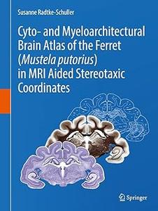
This stereotaxic atlas of the ferret brain provides detailed architectonic subdivisions of the cortical and subcortical areas in the ferret brain using high-quality histological material stained for cells and myelin together with in vivo magnetic resonance (MR) images of the same animal. The skull-related position of the ferret brain was established according to in vivo MRI and additional CT measurements of the skull. Functional denotations from published physiology and connectivity studies are mapped onto the atlas sections and onto the brain surface, together with the architectonic subdivisions. High-resolution MR images are provided at levels of the corresponding histology atlas plates with labels of the respective brain structures. The book is the first atlas of the ferret brain and the most detailed brain atlas of a carnivore available to date. It provides a common reference base to collect and compare data from any kind of research in the ferret brain.
Key Features
- Provides the first ferret brain atlas with detailed delineations of cortical and subcortical areas in frontal plane.
- Provides the most detailed brain atlas of a carnivore to date.
- Presents a stereotaxic atlas coordinate system derived from high-quality histological material and in vivo magnetic resonance (MR) images of the same animal.
- Covers the ferret brain from forebrain to spinal cord at intervals of 0.6 mm on 58 anterior-posterior levels with 5 plates each.
- Presents cell (Nissl) stained frontal sections (plate 1) and myelin stained sections (plate 2) in a stereotaxic frame.
- Provides detailed delineations of brain structures and their denomination on a Nissl stained background on a separate plate (3).
- Compiles abbreviations on plate 4, a plate that also displays the low resolution MRI of the atlas brain with the outlines of the Nissl sections in overlay.
- Displays high-resolution MR images at intervals of 0.15 mm from another animal with labeled brain structures as plate 5 corresponding to the anterior-posterior level of each atlas plate.
- Provides detailed references used for delineation of brain areas.
Target audience of the book:
The book addresses researchers and students in neurosciences who are interested in brain anatomy in general (e.g., for translational purposes/comparative aspects), particularly those who study the ferret as important animal model of growing interest in neurosciences.
Buy Premium From My Links To Get Resumable Support,Max Speed & Support Me
Links are Interchangeable - Single Extraction
Help Us Grow – Share, Support
We need your support to keep providing high-quality content and services. Here’s how you can help:
- Share Our Website on Social Media! 📱
Spread the word by sharing our website on your social media profiles. The more people who know about us, the better we can serve you with even more premium content! - Get a Premium Filehost Account from Website! 🚀
Tired of slow download speeds and waiting times? Upgrade to a Premium Filehost Account for faster downloads and priority access. Your purchase helps us maintain the site and continue providing excellent service.
Thank you for your continued support! Together, we can grow and improve the site for everyone. 🌐
Related News
-
{related-news}
Comments (0)

Information
Users of Guests are not allowed to comment this publication.
Search
Updates
Partner
» TutBB
» Byte
» Crawli
» Warezomen
» Warez-DDL
» Raidrush
» KATZCD
» Free Ebooks Library
Your Link Here ?
(Pagerank 4 or above)
» Byte
» Crawli
» Warezomen
» Warez-DDL
» Raidrush
» KATZCD
» Free Ebooks Library
Your Link Here ?
(Pagerank 4 or above)